Canine Hip Dysplasia
Canine Hip Dysplasia
The word dysplasia stems from the Greek words dys,
meaning “disordered” or “abnormal”, and plassein meaning “to form”.
Introduction to Dysplastic Hips
The expression hip dysplasia can be interpreted as the abnormal or faulty development of the hip. Abnormal development of the hip causes excessive wear of the joint cartilage during weight bearing, eventually leading to the development of arthritis, often called degenerative joint disease (DJD) or osteoarthritis (OA). The terms DJD, arthritis and osteoarthritis are used interchangeably.
Canine Hip Dysplasia (CHD) was first described in 1935 by Dr. Gerry B. Schnelle. Dr. Schnelle initially called it “bilateral congenital subluxation of the coxofemoral joint”. It was originally thought to be a rare condition but is now recognized as the most common orthopedic disease in dogs. This radiographic image is the first known example of CHD to be published in a scientific journal.
In 1966, Henricson, Norberg and Olsson refined the definition of CHD describing it as: “A varying degree of laxity of the hip joint permitting subluxation during early life, giving rise to varying degrees of shallow acetabulum and flattening of the femoral head, finally inevitably leading to osteoarthritis.”
Today, the general veterinary consensus is that hip dysplasia is a heritable disease manifested as hip joint laxity that leads to the development of OA. So a knowledge of hip joint laxity is key to predicting the ultimate development of the OA of CHD.
Canine Hip Dysplasia afflicts millions of dogs each year and can result in debilitating orthopedic disease of the hip. Many dogs will suffer from osteoarthritis, pain, and lameness, costing owners and breeders millions of dollars in veterinary care, shortened work longevity, and reduced performance. The occurrence of CHD is well documented in large and giant breed dogs, but there is also evidence that CHD is prevalent in many small and toy breeds as well as in cats.
Hip dysplasia is a disease of complex inheritance, meaning it is caused by many genes and its expression can be affected by multiple non-genetic factors. Veterinarians and dog breeders have attempted to eliminate CHD through selective breeding strategies based on scoring of the standard hip-extended radiograph, however, the reduction of CHD frequency in pure-breed dogs has been disappointing.
Fast facts about Canine Hip Dysplasia
- Canine hip dysplasia (CHD) is the most commonly inherited orthopaedic disease in dogs.
- CHD is a degenerative, developmental condition, leading to painful hip osteoarthritis, stiffness, and diminished quality of life.
- All dog breeds are affected by the disease: in some breeds more than 50% of dogs are afflicted.
- The disease is polygenic and multifactorial meaning that CHD expression is a product of both genes and environmental factors such as body weight and age.
- There is no medical or surgical cure for CHD.
- CHD is a major concern for working dog agencies, pet owners, dog breeders and veterinarians.
Development of Canine Hip Dysplasia
Canine hip dysplasia (CHD) is a developmental disease.
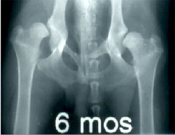
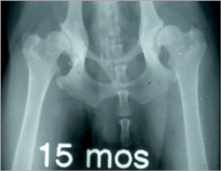
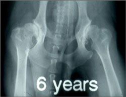
A developmental disease is not present at birth but develops with maturation and age. The series of radiographs below illustrate how a loose hip gradually develops osteoarthritis (OA).

At six months, this dog’s hips exhibit extreme laxity, but no OA.

At 15 months, laxity is accompanied by the development of “mild” to “moderate” OA: the femoral heads appear slightly “flattened”, the femoral necks are beginning to thicken and the acetabular rims are in the early stages of remodeling.

At six years, OA has progressed into a “severe” form, marked by extreme bony remodeling of the acetabular cups and the femoral head and necks. Cartilage has been eroded and bone is rubbing on bone.
Clinical Signs of Hip Dysplasia
Canine hip dysplasia (CHD) in its severest form can be diagnosed by clinical signs, but it usually requires radiographic evidence of hip joint laxity and/or the appearance of osteoarthritis (OA) to arrive at a definitive diagnosis.
There is an acute and a severe form of CHD. An affected dog may have one or any combination of the following clinical signs:
Severe (Acute) Form
- Presentation at five to 12 months of age,
- Overt pain, lameness, and functional deficits (low exercise tolerance, reluctance to climb stairs),
- Other signs: audible “click” when walking, increased intertrochanteric width (“points of hips” are wider than normal), thigh muscle atrophy.
Mild (Chronic) Form
- Clinical signs ranging from none to mild,
- Mild discomfort and stiffness in geriatric years,
- Possible pain and crepitus on range of motion.
Clinical signs by themselves do not necessarily mean that a dog has hip dysplasia, other conditions of the hip or knee can mimic CHD. A radiograph is essential for a more accurate assessment of a dog’s hip joint integrity.
Defining Hip Laxity
Hip joint laxity is the most important risk factor for the development of osteoarthritis.
In other words, the amount of laxity or looseness in a hip joint is related to the chance that a hip will develop OA: the looser the hip, the greater the risk. For this reason, it is important to understand the difference between passive and functional hip laxity.
- Passive hip laxity is subjectively scored or measured on a hip radiograph of a dog while under heavy sedation or anesthesia. The PennHIP method measures passive laxity.
- Functional hip laxity is the pathologic form of laxity occurring during normal weightbearing in dogs with dysplastic hips. Current hip screening methods cannot assess functional hip laxity.
- PennHIP research has shown that passive hip laxity is a clinically useful surrogate for functional hip laxity.
To see how PennHIP measures passive hip laxity, go to the Distraction Index – Measuring Laxity section.
Effects of Functional Laxity on Joint Mechanics
Under normal conditions, the sum of the forces on the joint are spread out over a large surface area of cartilage. When laxity (or subluxation) is present in the joint, the force applied by the surrounding muscles actually increases to compensate for the laxity (see middle figure). The sum of the forces exerted on the dysplastic hip is greater than the sum of forces exerted on the normal hip. In addition, the forces on the dysplastic hip are applied over a smaller surface area as shown at the contact point in the figure. The high joint contact stresses produce injury and ultimately result in the loss of delicate articular cartilage. Over time, functional hip laxity results in erosion of the femoral head and flattening of the acetabulum.
Osteoarthritis
OA causes pain and disability. OA affects all components of the synovial joint.
Osteoarthritis (OA): The Big Picture
- There is no cure for OA. There are palliative treatment options for OA.
- Non-surgical treatments include use of NSAIDS and nutraceuticals, the modification of nutrition, moderate exercise, and physical therapy.
- Surgery may be the only option for end-stage disease (ex., FHO, THR).
- Prevention is key in the fight against canine hip dysplasia (CHD) and OA:
- Accurate prediction of OA requires a reliable screening method implemented early in life.
- Genetic control and selective breeding based on a validated phenotype can reduce the severity of CHD and the development of OA in subsequent generations of animals.
- Environmental factors such as diet, activity level, and pharmaceuticals can influence the onset of OA.
- Preventive Surgery is also an option (eg, TPO, JPS). However, the safety and efficacy of preventive surgical procedures have not been adequately studied .
